| Kidney Cancer
Renal cell cancer is a disease in which
malignant cells form in tubules of the kidney.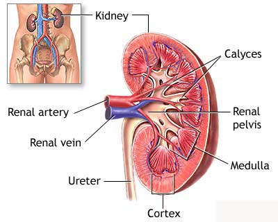
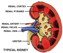
Renal cell cancer (also called kidney cancer or renal adenocarcinoma) is a
disease in which malignant (cancer) cells are found in the lining of tubules
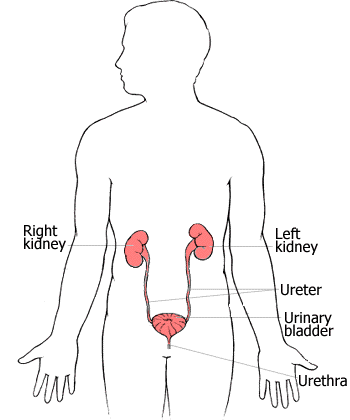
(very small tubes) in the kidney. There are 2 kidneys, one on each side of the
backbone, above the waist. The tiny tubules in the kidneys filter and clean the
blood, taking out waste products and making urine. The urine passes from each
kidney into the bladder through a long tube called a ureter. The bladder stores
the urine until it is passed from the body.
Cancer that starts in the ureters or the renal pelvis (the part of the kidney
that collects urine and drains it to the ureters) is different from renal cell
cancer.
Smoking and misuse of certain pain medicines can affect the risk of developing
renal cell cancer.
Risk factors include the following:
- Being a smoker
- Misusing certain pain
medicines, including over-the-counter pain medicines, for a long
time
- Having certain genetic conditions, such as von Hippel-Lindau
disease or hereditary papillary renal cell carcinoma
- Possible signs of renal cell cancer include blood in the urine and a lump in the
abdomen.
These and other symptoms may be caused by renal cell cancer or by other
conditions
There may be no symptoms in the early stages.
Symptoms may appear as the tumour grows. A doctor should be consulted if any of
the following problems occur:
- Blood in the urine
- A lump in the abdomen
- A pain in the side that
doesn't go away
- Loss of appetite
- Weight loss for no
known reason
- Anaemia
- Tests that examine the abdomen and kidneys are used to detect (find) and
diagnose renal cell cancer
The following tests and procedures may be
used:
Physical exam and history: An exam of the body to check general signs of
health, including checking for signs of disease, such as lumps or anything else
that seems unusual. A history of the patient’s health habits and past illnesses
and treatments will also be taken.
Blood chemistry studies: A procedure in
which a blood sample is checked to measure the amounts of certain substances
released into the blood by organs and tissues in the body. An unusual (higher or
lower than normal) amount of a substance can be a sign of disease in the organ
or tissue that produces it.
Urinalysis: A test to check the colour
of urine and its contents, such as sugar, protein, blood, and bacteria.
Liver function test: A procedure in
which a sample of blood is checked to measure the amounts of enzymes released
into it by the liver. An abnormal amount of an enzyme can be a sign that cancer
has spread to the liver. Certain conditions that are not cancer may also
increase liver enzyme levels.
Intravenous pyelogram (IVP): A series
of x-rays of the kidneys, ureters, and bladder to check for cancer. A contrast
dye is injected into a vein. As the contrast dye moves through the kidneys,
ureters, and bladder, x-rays are taken to see if there are any blockages.
Ultrasound: A procedure in which
high-energy sound waves (ultrasound) are bounced off internal tissues or organs
and make echoes. The echoes form a picture of body tissues called a sonogram.
CT scan (CAT scan): A procedure that
makes a series of detailed pictures of areas inside the body, taken from
different angles. The pictures are made by a computer linked to an x-ray
machine. A dye may be injected into a vein or swallowed to help the organs or
tissues show up more clearly. This procedure is also called computed tomography,
computerized tomography, or computerized axial tomography.
MRI (magnetic resonance imaging): A
procedure that uses a magnet, radio waves, and a computer to make a series of
detailed pictures of areas inside the body. This procedure is also called
nuclear magnetic resonance imaging (NMRI).
Biopsy: The removal of cells
or tissues so they can be viewed under a microscope to check for signs of
cancer. A thin needle is inserted into the tumour and a sample of tissue is
withdrawn. A pathologist then views the tissueunder a microscope to check for
cancer cells.
 The prognosis and treatment options
depend on the following: The prognosis and treatment options
depend on the following:
The stage of the disease.
The patient's age and general health.
After renal cell cancer has been diagnosed, tests are done to find out if cancer
cells have spread within the kidney or to other parts of the body.
The process used to find out if cancer has spread within the kidney or to other
parts of the body is called staging. The information gathered from the staging
process determines the stage of the disease. It is important to know the stage
in order to plan treatment. The following tests and procedures may be used in
the staging process:
CT scan (CAT scan): A procedure that makes a series of detailed pictures
of areas inside the body, taken from different angles. The pictures are made by
a computer linked to an x-ray machine. A dye may be injected into a vein or
swallowed to help the organs or tissues show up more clearly. This procedure is
also called computed tomography, computerized tomography, or computerized axial
tomography.
MRI (magnetic resonance imaging): A
procedure that uses a magnet, radio waves, and a computer to make a series of
detailed pictures of areas inside the body. This procedure is also called
nuclear magnetic resonance imaging (NMRI).
Chest x-ray: An x-ray of the organs and
bones inside the chest. An x-ray is a type of energy beam that can go through
the body and onto film, making a picture of areas inside the body.
Bone scan: A procedure to check if
there are rapidly dividing cells, such as cancer cells, in the bone. A very
small amount of radioactive material is injected into a vein and travels through
the bloodstream. The radioactive material collects in the bones and is detected
by a scanner.
The following stages are used for renal cell
cancer:
Stage I
In stage I, the tumour is no larger than 7 centimetres and is found in the
kidney only.
Stage II
In stage II, the tumour is larger than 7 centimetres and is found in the kidney
only.
Stage III
In stage III, cancer is found:
in the kidney and in 1 nearby lymph node; or
in an adrenal gland or in the layer of fatty tissue around the kidney, and may
be found in 1 nearby lymph node; or
in the main blood vessels of the kidney and may be found in 1 nearby lymph node.
Stage IV
In stage IV, cancer has spread:
beyond the layer of fatty tissue around the kidney and may be found in 1 nearby
lymph node; or to 2 or more nearby lymph nodes; or to other organs, such as the
bowel, pancreas, or lungs, and may be found in nearby lymph nodes.
Four types of standard treatment are used:
Surgery
Surgery to remove part or all of the kidney is often used to treat renal cell
cancer. The following types of surgery may be used:
Partial nephrectomy: A surgical procedure to remove the cancer within the kidney
and some of the tissue around it. A partial nephrectomy may be done to prevent
loss of kidney function when the other kidney is damaged or has already been
removed.
Simple nephrectomy: A surgical procedure to remove the kidney only.
Radical nephrectomy: A surgical procedure to remove the kidney, the adrenal
gland, surrounding tissue, and, usually, nearby lymph nodes.
A person can live with part of 1 working kidney, but if both kidneys are removed
or not working, the person will need dialysis (a procedure to clean the blood
using a machine outside of the body) or a kidney transplant (replacement with a
healthy donated kidney). A kidney transplant may be done when the disease is in
the kidney only and a donated kidney can be found. If the patient has to wait
for a donated kidney, other treatment is given as needed.
When surgery to remove the cancer is not possible, a treatment called arterial
embolization may be used to shrink the tumour. A small incision is made and a
catheter (thin tube) is inserted into the main blood vessel that flows to the
kidney. Small pieces of a special gelatine sponge are injected through the
catheter into the blood vessel. The sponges block the blood flow to the kidney
and prevent the cancer cells from getting oxygen and other substances they need
to grow.
Even if the doctor removes all the cancer that can be seen at the time of the
surgery, some patients may be given chemotherapy or radiation therapy after
surgery to kill any cancer cells that are left. Treatment given after the
surgery, to increase the chances of a cure, is called adjuvant therapy.
Radiation therapy
Radiation therapy is a cancer treatment that uses high-energy x-rays or other
types of radiation to kill cancer cells. There are 2 types of radiation therapy.
External radiation therapy uses a machine outside the body to send radiation
toward the cancer. Internal radiation therapy uses a radioactive substance
sealed in needles, seeds, wires, or catheters that are placed directly into or
near the cancer. The way the radiation therapy is given depends on the type and
stage of the cancer being treated.
Chemotherapy
Chemotherapy is a cancer treatment that uses drugs to stop the growth of cancer
cells, either by killing the cells or by stopping the cells from dividing. When
chemotherapy is taken by mouth or injected into a vein or muscle, the drugs
enter the bloodstream and can reach cancer cells throughout the body (systemic
chemotherapy). When chemotherapy is placed directly into the spinal column, a
body cavity such as the abdomen, or an organ, the drugs mainly affect cancer
cells in those areas. The way the chemotherapy is given depends on the type and
stage of the cancer being treated.
Biologic therapy
Biologic therapy is a treatment that uses the patient's immune system to fight
cancer. Substances made by the body or made in a laboratory are used to boost,
direct, or restore the body's natural defences against cancer. This type of
cancer treatment is also called biotherapy or immunotherapy.
Other types of treatment are being tested in clinical trials. These include the
following:
Stem cell transplantation
Stem cells (immature blood cells) are removed from the blood or bone marrow of a
donor and given to the patient through an infusion. These re-infused stem cells
grow into (and restore) the body's blood cells.
Treatment Options by Stage
Stage I Renal Cell Cancer
Standard treatment of stage I renal cell cancer may include the following:
Surgery (radical nephrectomy, simple nephrectomy, or partial nephrectomy).
Radiation therapy as palliative therapy to relieve symptoms in patients who
cannot have surgery.
Arterial embolization, as palliative therapy.
Stage II Renal Cell Cancer
Standard treatment of stage II renal cell cancer may include the following:
Surgery (radical nephrectomy or partial nephrectomy).
Surgery (nephrectomy), before or after radiation therapy.
Radiation therapy as palliative therapy to relieve symptoms in patients who
cannot have surgery.
Arterial embolization, as palliative therapy.
New treatments for stage II renal cell cancer are being studied in clinical
trials. Information about these and other ongoing clinical trials is available
from the NCI Cancer.gov Web site.
Stage III Renal Cell Cancer
Standard treatment of stage III renal cell cancer may include the following:
Surgery (radical nephrectomy). Blood vessels of the kidney and some lymph nodes
may also be removed.
Arterial embolization followed by surgery (radical nephrectomy).
Radiation therapy, as palliative treatment to relieve symptoms and improve the
quality of life.
Arterial embolization, as palliative therapy.
Surgery (nephrectomy), as palliative treatment.
Radiation therapy before or after surgery (radical nephrectomy).
One of the treatments being studied in clinical trials for stage III renal cell
cancer is biologic therapy following surgery.
Stage IV Renal Cell Cancer
Standard treatment of stage IV renal cell cancer may include the following:
Biologic therapy.
Radiation therapy as palliative treatment to relieve symptoms and improve the
quality of life.
Surgery (nephrectomy), as palliative treatment.
Surgery (radical nephrectomy, with or without removal of cancer from other areas
where it has spread).
New treatments for stage IV renal cell cancer are being studied in clinical
trials.
Treatment Options for Recurrent Renal Cell Cancer
Standard treatment of recurrent renal cell cancer may include the following:
Biologic therapy.
Radiation therapy as palliative treatment to relieve symptoms and improve the
quality of life.
Chemotherapy.
BACK
|


 Transitional cell carcinoma: Cancer that begins in cells in the innermost
tissue layer of the bladder. These cells are able to change shape depending on
whether the bladder is full or empty and may be stretched without breaking
apart. Most bladder cancers begin in the transitional cells.
Transitional cell carcinoma: Cancer that begins in cells in the innermost
tissue layer of the bladder. These cells are able to change shape depending on
whether the bladder is full or empty and may be stretched without breaking
apart. Most bladder cancers begin in the transitional cells.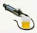 Urinalysis:
A test to check the colour of urine and its contents, such as sugar, protein,
blood, and bacteria.
Urinalysis:
A test to check the colour of urine and its contents, such as sugar, protein,
blood, and bacteria.  There
may be no symptoms of early cancer of the urethra. A doctor should be seen if
there is a lump or growth on the urethra, or pain, bleeding, or other difficulty
during urination.
There
may be no symptoms of early cancer of the urethra. A doctor should be seen if
there is a lump or growth on the urethra, or pain, bleeding, or other difficulty
during urination.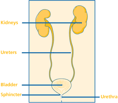
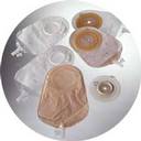 opening
(stoma) on the outside of the body. This is sometimes called an ostomy or urostomy. If a patient has an ostomy, a special
bag will need to be worn to collect urine. This special bag, which sticks to the
skin around the stoma with a special glue, can be thrown away after it is used.
This bag does not show under clothing, and most people take care of these bags
themselves. The doctor may also use part of the small intestine to make a new
storage pouch (a continent reservoir) inside the body where the urine can
collect. The patient would then need to use a tube (catheter) to drain the urine
through a stoma.
opening
(stoma) on the outside of the body. This is sometimes called an ostomy or urostomy. If a patient has an ostomy, a special
bag will need to be worn to collect urine. This special bag, which sticks to the
skin around the stoma with a special glue, can be thrown away after it is used.
This bag does not show under clothing, and most people take care of these bags
themselves. The doctor may also use part of the small intestine to make a new
storage pouch (a continent reservoir) inside the body where the urine can
collect. The patient would then need to use a tube (catheter) to drain the urine
through a stoma.